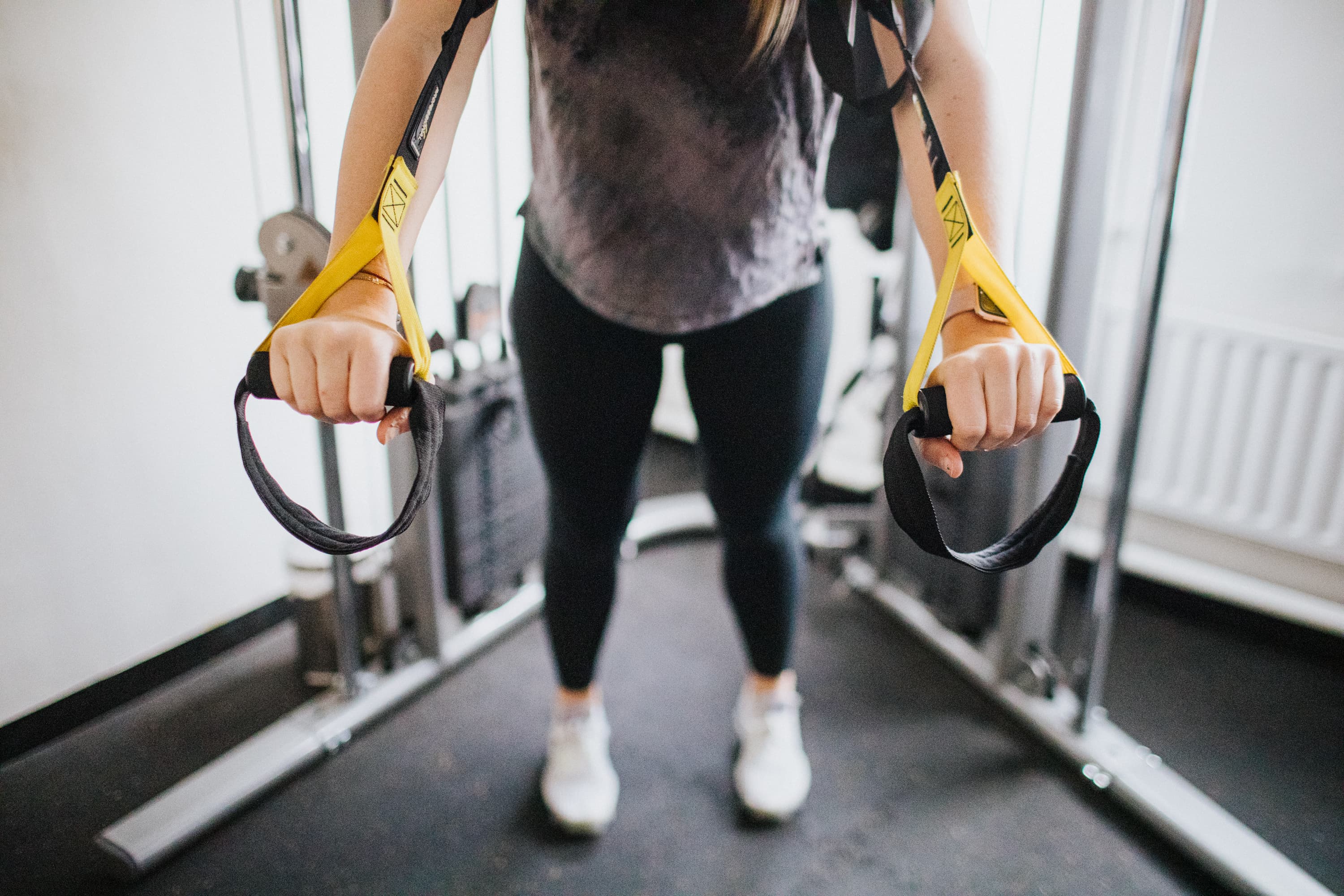
CG C071

Diagnostic Ultrasound Scans:

What is a diagnostic ultrasound scan?

A diagnostic ultrasound scan is an imaging technique that uses high-frequency sound waves to produce images of the body. It can be used to diagnose various musculoskeletal injuries, such as tendon tears, ligament sprains, and muscle strains. We commonly use it to diagnose the cause of shoulder pain, knee pain and elbow pain – as well as other areas.

How can ultrasound scans be used to diagnose musculoskeletal injuries?

Ultrasound scans are a highly effective tool for diagnosing musculoskeletal injuries. By sending sound waves through the tissues, it is possible to evaluate and diagnose soft tissue injuries, including tendon tears, ligament sprains, and muscle strains. Ultrasound images can show not only the injury itself but also the extent of the damage, helping to provide a more accurate diagnosis.

Benefits of using an ultrasound scan for diagnosis?

Using ultrasound to diagnose musculoskeletal injuries has many benefits. It is highly accurate, non-invasive, painless, and can be performed quickly and easily. Superficial joints, such as the shoulder joint, are easily seen with ultrasound. Ultrasound scans are also more cost-efficient than other diagnostic techniques such as X-rays and MRI scans.

Type of Musculoskeletal Injuries Diagnosed with Ultrasound Scans?

Ultrasound scans can be used to diagnose a wide range of musculoskeletal injuries, including sports injuries, tendon tears, ligament sprains, and muscle strains. Injuries including rotator cuff tears (shoulder pain), patella tendonitis (knee pain) and tennis elbow (elbow pain) are all easily diagnosed with Ultrasound.

Ultrasound images can show the extent of the injury as well as any associated inflammation or fluid accumulation. The results of the scan can then be used to determine the course of treatment for the injury.

Sports Injuries:

Ultrasound scans can be used to diagnose a variety of sports injuries, including ankle sprains, knee ligament tears, shoulder pain, rotator cuff tendinitis, non-specific knee pain and hamstring strains.

Tendon Injuries:

Ultrasound scans can be used to diagnose a variety of tendon injuries, including tears, tendinitis, and ruptures to the Rotator Cuff tendons, Achilles Tendon, Wrist and Hand tendons and Patellar tendon.

Ligament and Joint Injuries:

Ultrasound scans can be used to diagnose a variety of ligament tears, including MCL injuries (knee ligament injury), ankle ligament injuries, shoulder labral tears, and meniscus and knee cartilage tears (knee joint injury). Ultrasound images can show the extent of the injury as well as any associated inflammation.

Muscle Injuries:

Ultrasound scans can be used to diagnose a variety of muscle injuries, including strains, contusions, and tears. Ultrasound images can show the extent of the injury to categorise it and predict the recovery time.

Knee Pain:

Ultrasound scans can be used to diagnose a variety of knee joint pain, including patellar tendonitis, meniscus tears and other ligament injuries. Ultrasound images can help show the extent of the knee injury as well as any associated inflammation or fluid accumulation. Ultrasound is also highly effective for diagnosing non-specific knee problems and problems suffered by young adults such as Osgood-Schlatter’s.

Shoulder Pain:

Ultrasound scans can be used to diagnose a variety of shoulder pain, including rotator cuff tendonitis, bursitis and labral tears. Causes of severe pain in the shoulder, such as calcification and frozen shoulder, can also be picked up. Ultrasound images provide detailed information on the structure and function of the shoulder joint and can detect even small changes in tissue composition that may cause a shoulder problem.

Rotator Cuff Tendons:

A Rotator Cuff injury is the most common complaint that causes shoulder problems. Many people feel pain during exercise or for a few days after, and often refers pain into the arm, the back of the shoulder area or into the front of the joint.

Ultrasound scans can be used to diagnose rotator cuff tendon injuries, such as tears, tendinitis and ruptures. Ultrasound images provide detailed information on the structure of the rotator cuff tendons and can detect even small changes in tissue composition, known as tendinopathy.

Rotator cuff injuries are easily diagnosed with ultrasound scans because the tendons are located close to the surface of the skin and can be seen clearly in an ultrasound image. Ultrasound images provide detailed information on the structure of these tendons, allowing for accurate diagnosis and treatment.

Preparing for an Ultrasound Scan:

The scan will usually accompany a series of questions about how the pain started and a physical exam to ensure the area is thoroughly assessed. It is important to wear loose-fitting clothing that can be easily removed. It is recommended that you drink plenty of water prior to the scan in order to ensure that your muscles are hydrated and relaxed.

What to expect during the scan:

During the scan, a technician will use a small ultrasound probe to transmit sound through your body. The sound waves will bounce off structures within your body and be picked up by the probe, creating an image on the ultrasound device screen that can then be used for diagnosis. The scan can also pick up blood flow to the affected tissue.

Ultrasound scans are completely painless and non-invasive and typically take around 30 minutes to complete.

Post-scan instructions and guidelines:

Following an ultrasound scan, it is important to follow any instructions provided by your Physiotherapist. This may include the use of cold or heat therapy, rest, physiotherapy, and medication. You will receive a report detailing exactly what was seen in the assessment, for your records.

Potential follow-up treatment protocols:

Depending on the results of your ultrasound scan and the severity of your injury, there are a variety of treatment protocols that may be recommended. These can include physiotherapy, rest, taking analgesics to provide relief or anti-inflammatory medication, and in some cases orthopaedic surgery.

There are other physical therapy options such as Shockwave Therapy, Strength & Conditioning and Dry Needling – depending on the diagnosis.

Your physiotherapist will discuss these options with you and help to decide which is most appropriate for your condition.

Summary:

Ultrasound scans are a valuable tool to diagnose musculoskeletal injuries, including sports injuries, tendon tears, ligament sprains and muscle strains – as well as unclear causes of shoulder pain, knee pain and elbow pain.

Diagnose and Relieve Pain with Carter & George:

At Carter & George Physiotherapy, we provide diagnostic ultrasound scans to help diagnose and manage musculoskeletal injuries, alongside other modern technological healthcare options. Our experienced team of physiotherapists are highly trained in the use of ultrasound and can provide you with an accurate diagnosis and effective treatment plan for your injury. We will use the information to strategize the most effective and efficient treatment options for you to relieve pain, reduce inflammation and get you back to your best.

Please note, we use diagnostic ultrasound scans for musculoskeletal injuries only and do not assess hernias, lumps or bumps.

Due to the depth ultrasound can get to, it is not used to examine spinal injuries. Small broken bone and fractures can be seen, but other imaging tests such as X rays are more effective for these medical conditions.

Rheumatoid Arthritis can often cause similar symptoms to musculoskeletal pain, however a blood test may be required to confirm this diagnosis.

If you require onward referral for other tests such as MRI, CT imaging studies or X-Ray, your Physiotherapist will be able to assist.